

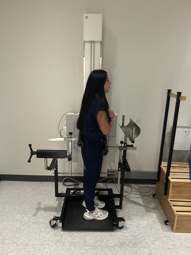
When clinicians use flexion and extension X-rays to evaluate the lumbar spine, the objective is to see how the spine behaves during movement. Unlike a static image, a dynamic examination can provide information about changes in alignment, angulation, and translation as the patient moves between positions. But obtaining a meaningful dynamic image depends on more than the X-ray equipment itself. The way a patient moves during the examination can also influence what the resulting images show.
Andrew Maxwell has focused on this often overlooked aspect of lumbar imaging, particularly the difficulty of isolating movement through the lumbar spine during flexion and extension examinations. Patients may naturally guard against movement because of discomfort, or they may compensate by moving through the hips rather than producing the intended motion through the lumbar region. When that happens, the examination may become less consistent from one patient or study to another.
The issue is important because flexion and extension imaging is often used when clinicians are interested in spinal movement and potential instability. If the movement being captured does not adequately represent the lumbar spine, the resulting image may provide a less complete picture of what is occurring.
This is the problem Maxwell has sought to address through Flexmax, an accessory designed for use in the X-ray room during lumbar flexion and extension examinations. Rather than changing the underlying imaging technology, the concept focuses on patient positioning and controlling some of the physical variables that can influence a dynamic examination.
The principle is relatively straightforward. If the purpose of flexion and extension imaging is to evaluate lumbar movement, then reducing unintended movement elsewhere in the body may help make that movement easier to assess. Flexmax is designed to help limit hip hinging and compensatory movement while supporting the intended motion of the lumbar spine.
“Flexion and extension imaging is intended to show how the spine moves, so the quality of that movement matters,” Maxwell explains. “If the patient is guarding or moving primarily through the hips, the examination may not represent the lumbar motion that the clinician is trying to evaluate.”
Patient movement is not necessarily a technical problem that can be solved through imaging equipment alone. Dynamic examinations involve interaction between the patient, the technologist, positioning, instructions, and the physical mechanics of movement. Even when the same imaging protocol is followed, differences in how a patient performs the required motion can introduce variability into the examination.
That variability becomes especially relevant when clinicians are looking for relatively subtle changes in lumbar alignment. Translation and angulation can be important considerations when assessing instability, and identifying those changes depends on obtaining images that accurately represent the movement being evaluated.
According to Maxwell, one of the goals behind Flexmax is to reduce that variability. He believes that by helping isolate lumbar movement and limiting compensatory hip movement, the accessory could improve the consistency of flexion and extension examinations and make instability easier to identify.
Maxwell has described the potential impact in significant terms, suggesting that the use of Flexmax could double the detection of lumbar angulation and translation instability while reducing false-negative examinations. Those claims ultimately require evaluation through clinical use and appropriate evidence, but they point to the underlying question behind the device: how much does examination technique influence the information clinicians receive from a dynamic X-ray?
That question extends beyond Flexmax itself. Medical imaging is often discussed primarily in terms of scanners, detectors, resolution, and other technological capabilities. Yet the quality of an examination can also depend on factors that occur before an image is captured. Patient positioning and movement are part of that process, particularly when the purpose of the examination is to observe structures in motion.
For lumbar flexion and extension studies, this creates a practical challenge. The lumbar spine does not move in isolation from the rest of the body. The hips and pelvis are naturally involved in movement, and patients may alter their technique because of pain, discomfort, apprehension, or simple differences in how they interpret instructions. A dynamic image can therefore reflect a combination of movements rather than exclusively the movement clinicians intended to evaluate.
This is where Maxwell’s approach takes a different direction. Instead of treating positioning as a secondary consideration, he has focused on it as a potential source of variability within the examination itself.
Flexmax is intended to function as an accessory rather than a replacement for existing X-ray systems or professional clinical judgment. Its purpose is to assist with the physical setup of the examination and help create conditions in which lumbar flexion and extension can be captured more consistently.
The broader significance of this approach lies in its focus on reproducibility. When clinicians compare flexion and extension images, they are attempting to understand what changes between positions. If the patient’s movement differs substantially from one examination to another, distinguishing genuine spinal movement from differences in technique can become more difficult.
Improving consistency does not eliminate every variable involved in dynamic imaging, nor does it determine how a clinician should interpret an examination. What it can potentially do is address one part of the process: how the patient’s movement is controlled and represented in the resulting images.
For Maxwell, that distinction is central to the thinking behind Flexmax.
“When clinicians are assessing instability, they need the images to reflect the movement they are trying to evaluate,” he says. “Reducing variability can help make the examination more consistent.”
The concept also reflects a broader shift in how medical imaging is understood. Static images remain fundamental to diagnostic care, but movement can provide another layer of information in situations where the relationship between structures changes with position. Dynamic imaging therefore places additional importance on how an examination is performed, because the image is intended to capture not just anatomy, but anatomy in motion.
As interest in motion-based imaging continues, practical questions around positioning and examination technique are likely to remain relevant. The technology used to capture an image may be highly sophisticated, but the quality of the information can still depend on whether the physical conditions of the examination allow the intended movement to be observed.
Maxwell’s work with Flexmax centers on that principle. By addressing hip compensation and guarding during lumbar flexion and extension X-rays, the device is designed to tackle a specific challenge that can otherwise be easy to overlook.
Whether that approach produces the level of improvement Maxwell anticipates will ultimately depend on clinical experience, further evaluation, and how imaging professionals assess its performance in practice. But the underlying issue is broader than any individual product.
A dynamic X-ray is only as useful as the movement it captures. For clinicians attempting to understand lumbar instability, that makes the mechanics of the examination an important part of the imaging process itself.
By drawing attention to patient positioning, movement variability, and the difficulty of isolating lumbar motion, Maxwell’s work raises a practical question for the field: when the objective is to see how the spine moves, should greater attention be given to controlling how that movement is produced?
Media Contact:
Name: Andrew Maxwell
Email: [email protected]